Digestive Diseases and Sciences

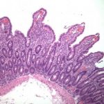
Celiac Disease Pathogenesis III 300×288
Treatment and ResponseTreatment data were available in 32 of 33 (97%) cases, and clinical response was documented in 30. Of 30, 24 (80%) showed a clear clinical response to antibiotic therapy, 2 of 30 (7%) no clinical response or further deterioration, and 4 of 30 (13%) equivocal clinical response after median follow-up of 14 months (IQR 2–39). Two of the four cases with equivocal clinical response were CNS disease treated with intravenous (IV) ceftriaxone and either oral trimetho-prim–sulfamethoxazole (TMP-SMX) or ceftibuten, and the two other cases were classic WD treated with ceftriaxone and TMP-SMX. The majority, 23 of 32 (72%), received a combination of IV ceftriaxone and oral TMP-SMX. TMP-SMX monotherapy was used in 7 of 32 (25%), minocycline monotherapy in 1 of 32 (4%), and 1 patient deferred ther-apy. Of the MCR cohort, 4 of 28 cases had serum levels of antibiotics followed.Lack of response to therapy or further deterioration occurred in two patients, one with classic WD and the other in the patient that deferred therapy. The lack of response in the classic WD case may represent immune reconstitution inflammatory syndrome (IRIS) as opposed to treatment failure, due to the initial response to pred-nisone; however, there was no confirmation of successful treatment through a negative tissue PCR, which is required for diagnosis of IRIS [31].All cases treated with T
Digestive Diseases and Sciences
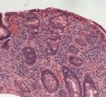
Celiac Disease Pathogenesis III 300×288
Digestive Diseases and Sciences
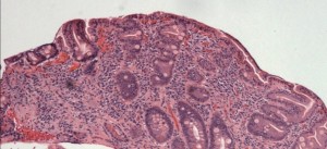
Celiac Disease Pathogenesis III 300×288
Treatment and ResponseTreatment data were available in 32 of 33 (97%) cases, and clinical response was documented in 30. Of 30, 24 (80%) showed a clear clinical response to antibiotic therapy, 2 of 30 (7%) no clinical response or further deterioration, and 4 of 30 (13%) equivocal clinical response after median follow-up of 14 months (IQR 2–39). Two of the four cases with equivocal clinical response were CNS disease treated with intravenous (IV) ceftriaxone and either oral trimetho-prim–sulfamethoxazole (TMP-SMX) or ceftibuten, and the two other cases were classic WD treated with ceftriaxone and TMP-SMX. The majority, 23 of 32 (72%), received a combination of IV ceftriaxone and oral TMP-SMX. TMP-SMX monotherapy was used in 7 of 32 (25%), minocycline monotherapy in 1 of 32 (4%), and 1 patient deferred ther-apy. Of the MCR cohort, 4 of 28 cases had serum levels of antibiotics followed.Lack of response to therapy or further deterioration occurred in two patients, one with classic WD and the other in the patient that deferred therapy. The lack of response in the classic WD case may represent immune reconstitution inflammatory syndrome (IRIS) as opposed to treatment failure, due to the initial response to pred-nisone; however, there was no confirmation of successful treatment through a negative tissue PCR, which is required for diagnosis of IRIS [31].

Celiac Disease Definition and Clinical Manifestations Dermatitis Herpetiformis 300×200

Celiac Disease Epidemiology poorly responsive II 300×147
Digestive Diseases and Sciences

Celiac Disease Diagnosis video capsule 300×300
Treatment and ResponseTreatment data were available in 32 of 33 (97%) cases, and clinical response was documented in 30. Of 30, 24 (80%) showed a clear clinical response to antibiotic therapy, 2 of 30 (7%) no clinical response or further deterioration, and 4 of 30 (13%) equivocal clinical response after median follow-up of 14 months (IQR 2–39). Two of the four cases with equivocal clinical response were CNS disease treated with intravenous (IV) ceftriaxone and either oral trimetho-prim–sulfamethoxazole (TMP-SMX) or ceftibuten, and the two other cases were classic WD treated with ceftriaxone and TMP-SMX. The majority, 23 of 32 (72%), received a combination of IV ceftriaxone and oral TMP-SMX. TMP-SMX monotherapy was used in 7 of 32 (25%), minocycline monotherapy in 1 of 32 (4%), and 1 patient deferred ther-apy. Of the MCR cohort, 4 of 28 cases had serum levels of antibiotics followed.Lack of response to therapy or further deterioration occurred in two patients, one with classic WD and the other in the patient that deferred therapy. The lack of response in the classic WD case may represent immune reconstitution inflammatory syndrome (IRIS) as opposed to treatment failure, due to the initial response to pred-nisone; however, there was no confirmation of successful treatment through a negative tissue PCR, which is required for diagnosis of IRIS [31].All cases treated with T
-
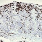
- Celiac Disease Pathogenesis III 300×288
-
- Celiac Disease Epidemiology poorly responsive 300×137
-
- Celiac Disease Epidemiology poorly responsive II 300×147
-
- Celiac Disease Pathogenesis II 300×225
